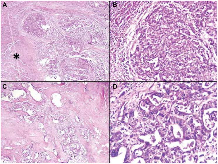
Figure 4.(A) Histological section demonstrating a poorly differentiated hepatocellular carcinoma component, characterized by tumor cells arranged in solid sheets, (Hematoxylin and eosin, × 100). (B) Histological section showing a poorly differentiated component of hepatocellular carcinoma, characterized by tumor cells arranged in solid sheets, displaying moderate nuclear atypia, (Hematoxylin and eosin, × 200). (C) Histopathological section showing a focus of cholangiocarcinoma, characterized by glandular structures of variable size lined by atypical malignant epithelial cells, surrounded by a prominent desmoplastic stroma, (Hematoxylin and eosin, × 100). (D) Histopathological section demonstrating a component of cholangiocarcinoma, characterized by closely apposed glandular structures composed of malignant epithelial cells embedded in a dense desmoplastic stroma, (Hematoxylin and eosin, × 400).