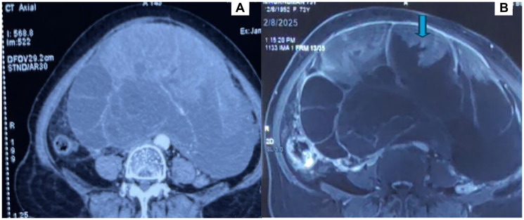
Figure 1.Radiological imaging of the abdominal lesion(A) CT axial view showing a large, lobulated, multiloculated solid cystic lesion in abdomen. (B) MRI T2 Image showing a large, lobulated, multiloculated solid cystic lesion in abdomen with hyperintense solid components (arrow).